SOLUTION Muscle diagrams Biology Diagrams
SOLUTION Muscle diagrams Biology Diagrams Learn about the muscular system, which is responsible for the movement of the human body. Explore detailed 3D anatomical illustrations of the skeletal, cardiovascular, digestive, endocrine, nervous, respiratory, immune, urinary, female reproductive, male reproductive, and integumentary systems. Learn about the three types of muscle tissue and the anatomy and function of skeletal muscles. Explore the 3D models of the muscular system and the naming of skeletal muscles.

Learn about the muscular system, a set of tissues that can change shape and produce force. See diagrams of skeletal, visceral and cardiac muscles, and how they work with the nervous system.
Interactive Guide to the Muscular System Biology Diagrams
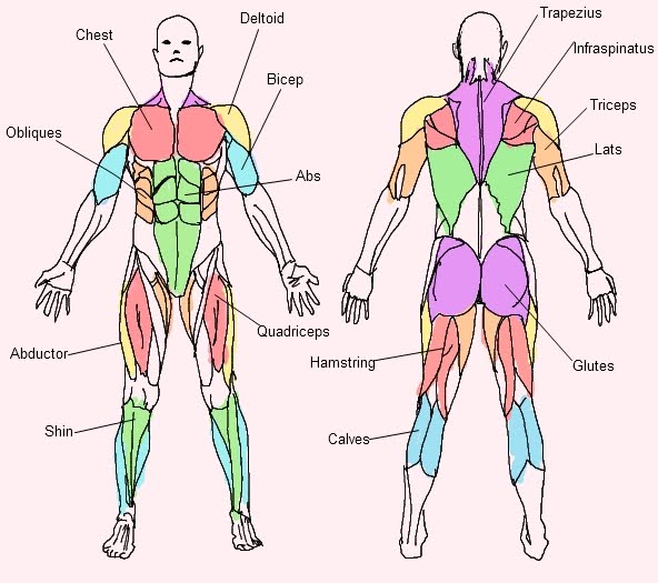
Muscular System Diagram. Here is the diagram of the human muscular system: Figure 5: Muscle chart showing the muscular system labeled where most muscles of the body are labeled in the form of a map of muscles. Image Credit: Wikimedia. Muscular System Physiology. Find out about the functions of the muscle tissues below. Learn the origins, insertions, innervations, and functions of all 600+ muscles in the body with high-quality illustrations. Download free or buy muscle charts for upper limb, lower limb, head and neck, and trunk wall in English or Latin terminology. Learn about the anatomy and physiology of muscles with Osmosis High-Yield Notes. See diagrams and illustrations of muscle fibers, sliding filament model, neuromuscular junction, and more.
